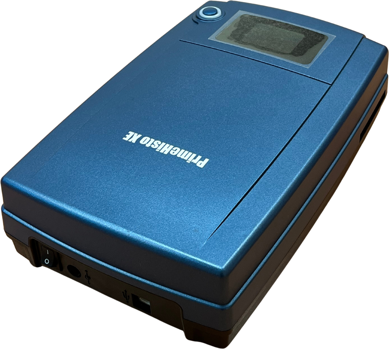
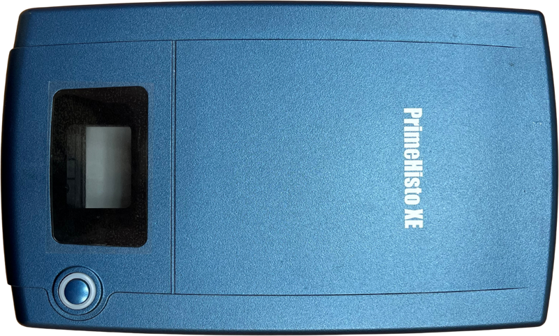
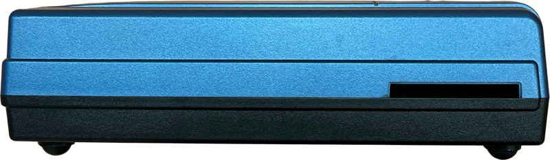

The PrimeHisto XE bridges the gap between traditional microscopy and digital workflows. It offers unmatched detail, real color accuracy, and powerful software tools in a compact, reliable package. Whether you’re documenting delicate histological preparations or building a digital archive for teaching and review, this scanner is optimized to deliver the clarity and consistency your work demands.
Key Features & Benefits
10,000 dpi resolution
Capture fine structural details in your tissue slides. Every cellular boundary, stain gradient, and morphological nuance is recorded with sharpness and accuracy. (Pacific Image Electronics Co., Ltd)True color reproduction with a linear CCD sensor
Unlike area sensors that interpolate color, the PrimeHisto XE uses a True Color Linear CCD for direct RGB capture, preserving the actual stain colors and intensities in your slides. (Pacific Image Electronics Co., Ltd)Stable slide holding system
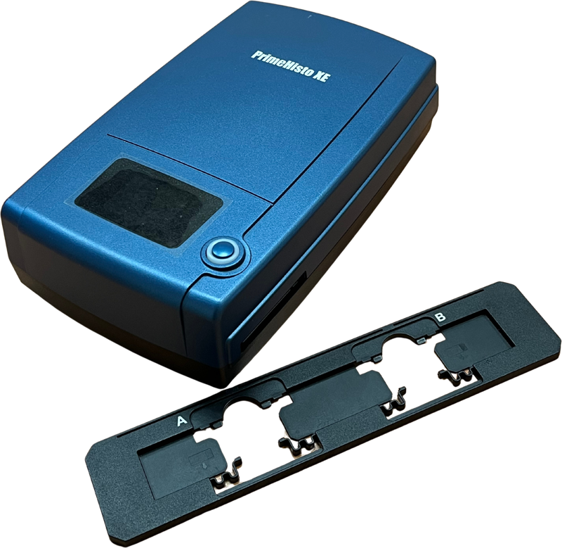
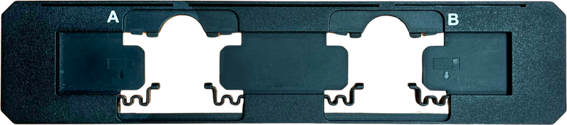
Includes a dual-slide holder with secure alignment strips, ensuring that slides remain firmly positioned during scanning. (Pacific Image Electronics Co., Ltd)Broad scanning range to support microscope integration
The scanner allows observation from about 10 µm up to 1 cm, making it easier to locate regions of interest and integrate with your microscope workflows. (Pacific Image Electronics Co., Ltd)Rich software support with HistoView
Bundled with Pacific Image’s HistoView software, you gain access to features like gamma curve adjustment, fine control over brightness, contrast, saturation, and more—designed specifically for histological image processing. (Pacific Image Electronics Co., Ltd)Flexible compatibility
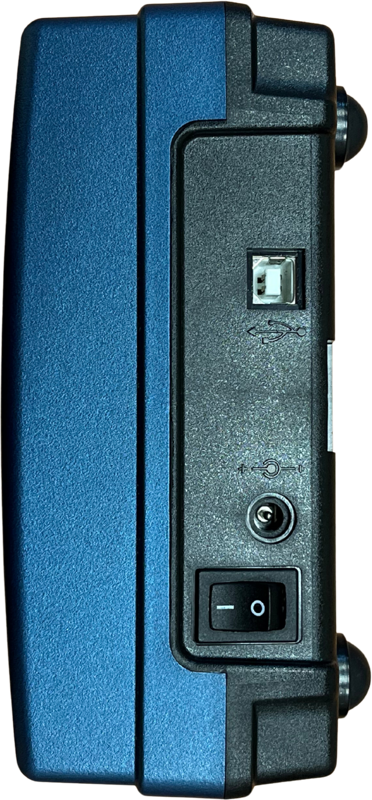
Works on both Windows (7 through 11) and macOS 10.13+ systems. (Pacific Image Electronics Co., Ltd)
USB 2.0 connectivity, with recommended host hardware of minimum 4 GB RAM (8 GB preferred). (Pacific Image Electronics Co., Ltd)
Specifications
| Scanning Media | Microscope slide |
| Resolution | Up to 10,000 dpi |
| Dynamic Range | 3.9 |
| Light Source | White LED (transmission) |
| Sensor | Linear array color CCD |
| Data Mode | 48-bit (color), 16-bit (grayscale) |
| Scan Area | 24.3 mm × 36.5 mm |
| File Formats | TIFF, BMP, JPEG |
| Power Supply | AC 100–240 V → 12 V DC, 1.5 A |
| Dimensions (L×W×H) | Approx. 275 × 167 × 80 mm |
| Weight | 2.1 kg |
| OS Support | Windows 7/8/10/11, macOS 10.13+ |
| Recommended Requirements | ≥4 GB RAM, 50 GB free disk space (Pacific Image Electronics Co., Ltd) |
Ideal Use Cases
Academic and research labs conducting histological or pathological examinations
Medical / clinical labs requiring digital archiving of slides
Teaching institutions where students learn tissue morphology and staining
Any environment needing high-fidelity digitization of microscope slides